Building such as houses have a frame that supports the roof and keeps the walls upright. Your body also has a supporting frame- a system of bones known as your skeleton.
Attached to this skeleton are your muscles. Together your bones and muscles form your skeletal/muscular system, which allows you to stand upright and to move about. This system also gives you shape and protects important organs such as your heart and brain.
Your skeleton is your body’s frame. It is made up of bones. the skeletons has three main functions:
- supporting your soft tissues. Without aa skeleton your skin and the organs inside your body would collapse to resemble a jellyfish stranded on the beach.
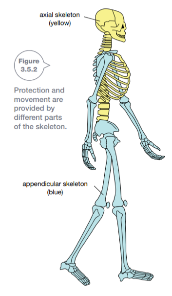
- Protecting your organs. This is the main role of your axial skeleton which is made up of 80 bones. It includes your skull (which protects your brain), vertebrae (which protect your spinal cord), and ribs and sternum or breastbone (which protect your lungs and heart).
- allowing movement. This is the main role of your appendicular skeleton, which is constructed from 126 bones. It includes your pelvis, shoulder blades and collarbones (clavicles), and the bones in your arms and legs.
Figure 3.5.2 shows your axial skeleton (shaded in yellow) and your appendicular skeleton (shaded in blue). Joints within the skeleton allow your body to move
Bones
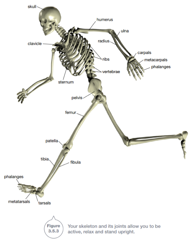
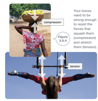
The main bones of your body are shown in Figure 3.5.3. Bones need to be strong so that they do not snap or crumble during normal activity. Compression forces squash bones and tension forces stretch them. Your leg bones experience compression forces when you are standing. The neck is usually under compression too, especially when carrying a load on the head, like the woman in Figure 3.5.4. Your arms experience tension forces whenever you carry shopping bags or when you hang off a bar like the gymnast in Figure 3.5.4.
Bones also need to be slightly elastic. They must be able to flex and twist a little and then return to their original shape and size when the force is removed from them.

Bones must also be light enough for your muscles to move. The strength and lightness of bones comes from their structure. The structure of a typical bone is shown in Figure 3.5.5. Bone tissue has two different forms.
- Compact bone makes up the outer layer of the bone. Compact bone is dense and heavy and gives the bone much of its strength.
- Spongy bone resembles honeycomb. It is light yet provides a strong inner structure to the bone. Gaps in spongy bone are filled with a fatty, jelly-like material called bone marrow. Red blood cells and platelets are made in bone marrow along with some white blood cells.
Bones are living tissues. They contain living cells that are surrounded by calcium phosphate, a substance that makes the bone hard. They also contain collagen, a material that provides elasticity. Blood vessels supply bone with the nutrients it requires. Bones are living, and hence able to repair themselves and replace worn-out cells.
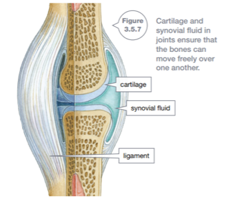
A joint is a place in the body where two bones come together. Most joints allow movement, and the type of joint determines the range of movement of the bones. Bone moving against bone would cause pain and the bone would wear down.
To protect your bones from wear, joints have:
- cartilage, a smooth and slippery material that covers the ends of bones that are moving against each other. It helps to cushion the impact of movement.
- synovial fluid, which lubricates the bone ends and allows them to slide over each other free
Types of joints
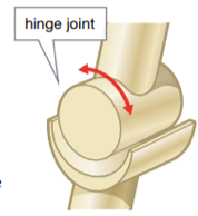
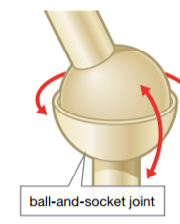
When you are sitting on a chair, your lower leg can move only forwards or backwards. The joint at the knee does not allow the lower leg to move from side to side. Using the hip joint, you can move your leg to make a complete circle with your toes. This is because your hip and knee are different types of joints.
In a hinge joint, the ends of the bones are shaped so that movement is allowed in only one plane-backwards and forwards hinge join like the hinge of a door. There is a hinge joint between the bone of the upper arm (humerus) and the larger bone of the lower arm (ulna). Other examples are the knee joint and joints in your fingers .
In a ball-and-socket joint, one bone has a ball-shaped surface that fits into a cup- shaped socket in the other bone. The bone with the ball at its end is able to complete all types of movement. You have ball-and-socket joints in your hips and shoulders.
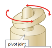
A pivot joint moves by a ring-shaped bone turning around another bone shaped like a finger. A pivot joint allows a wide range of movement. The joint at the base of your skull that allows you to tum your head is a pivot joint.
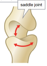
A saddle joint allows movement in two directions but does not allow the same amount of movement as a ball and socket joint. Saddle joints are found in the ankle and base of the thumb.
Ligaments
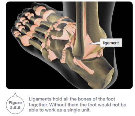
Ligaments are bands of tough, flexible tissue that hold the bones in a joint together. Ligaments prevent the bones of the joint from moving too far apart. Figure 3.5.8 shows the ligaments in the foot.
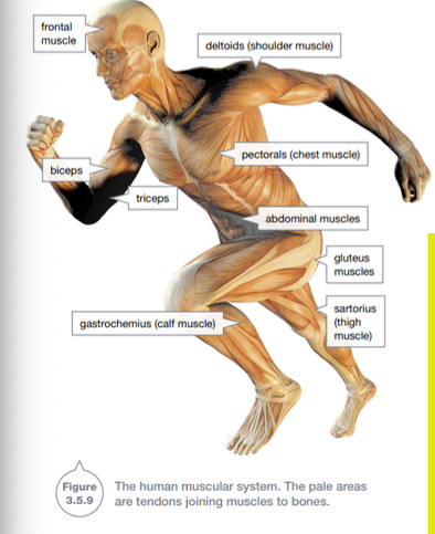
It is your muscles that move bones. Muscles are tissues that are able to contract (shorten) and be stretched. You have 640 muscles, regardless of whether you are a couch potato or a body builder. Some of the main muscles are shown in Figure 3.5.9. Tendons are tissues that attach muscles to bones and hold the muscles in position. When activated, muscles contract, becoming fatter and shorter. They pull on the bones they are attached to, causing them to move.
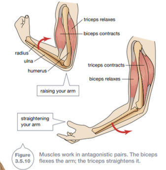
Muscles can only pull.They cannot push. Therefore, another muscle is required to return the bone to its original position. For this reason, your muscles are arranged in antagonistic pairs. Antagonistic means that they work in opposition to each other. The biceps and triceps are antagonistic muscles in your upper arm. The biceps contracts when activated, pulling your forearm up. The biceps then relaxes.
To lower the forearm, the triceps contracts. The relaxed biceps is stretched back to its original shape and the arm is straightened. This action is shown in Figure 3.5. 10.


